

Multimodal panoramic scanning single-cell LMD system
Main products


contactus
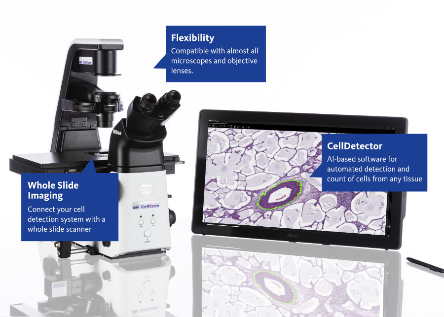
MMI CellCut and CellScan laser microdissection system: scan the whole slide, identify targets on the panoramic image, then cut and collect them on the same inverted research microscope. Authorized distributor in China: Oriental Horizon Technology Co Ltd (Beijing).
System overview
This configuration combines two systems from Molecular Machines & Industries (MMI, Germany) on one inverted research microscope: the CellScan multimodal panoramic scanning system and the CellCut laser microdissection system. The default host microscopes are the Nikon Ti2-E, EVIDENT IX83 / IX85, and ZEISS Axio Observer 7.
CellScan converts an entire slide into a high-resolution digital image. Targets are then outlined on that image and passed to CellCut on the same microscope, so that positional information remains in the coordinate system of the original slide. The configuration serves spatial multi-omics and related life-science research.

Figure. MMI CellCut and CellScan on one inverted research microscope.
Scan, identify, cut: the same-microscope workflow
- Scan the whole slide to produce a high-resolution digital image.
- Identify and outline targets on the image. Outlining can be performed manually, assisted by the optional CellDetector automated target recognition software, or based on external contours imported with the optional ShapeImport data import software.
- On the same inverted research microscope, CellCut performs laser microdissection with top-side collection, and the collected material can be checked visually on the tube cap.
- Positional information is retained in the coordinate system of the original slide, which facilitates comparison with downstream omics results.

Figure. Scan, outline and cut on one optical platform.
CellScan: panoramic scanning
- Defined by the manufacturer as 5D whole-slide imaging: XY scanning, Z-stack, time-lapse imaging, and multicolor fluorescence.
- Sample formats include standard slides, membrane slides for subsequent laser microdissection, and live cells in culture dishes. A stage incubation environment for time-lapse imaging can be configured according to the method; environmental control equipment is not part of the standard system.
- Images are stored in the open BigTIFF format and support remote viewing and annotation.
CellCut: laser microdissection
- Ultraviolet laser cutting of target regions, single cells, or subcellular structures in tissue sections. The laser position remains fixed while the stage moves along the outlined path, which keeps the focal plane stable.
- Top-side adhesive collection with CapSure / CapLift caps; after cutting, retention of the sample can be confirmed visually on the cap. Gravity-drop or catapult collection is not used.
- Suitable samples include formalin-fixed paraffin-embedded (FFPE) sections, fresh frozen sections, smears, cytospin preparations, plant tissue, and live cells. For live cells the laser enters from below, so the culture medium does not need to be removed.
- MMI CellTools software is used for outlining and recording.

Figure. CellCut and CellScan system detail.
Typical applications
- Spatial multi-omics and other studies that require whole-slide records with in situ coordinates and targeted sampling.
- High-resolution whole-slide digitization of tissue sections and membrane slides followed by laser microdissection of selected regions.
- Research workflows that link panoramic imaging directly to laser microdissection on the same microscope.
Related systems
For the individual systems, see the CellScan and CellCut product pages. Capillary isolation of cells in suspension and of adherent cells is performed with CellEctor; infrared optical tweezers are provided by CellManipulator.
Oriental Horizon Technology Co Ltd (Beijing) / 北京欧浩科技有限公司
Authorized distributor in China for Molecular Machines & Industries (MMI, Germany)
Address: Room 2703, Building A, Times Fortune Plaza, No. 1 Hangfeng Road, Fengtai District, Beijing
Demonstrations and evaluations: Ms. Zhang · judy.cheung@ohhktech.com · Mobile +86 135 1102 9964 · Landline 010 81311551
Manufacturer: molecular-machines.com · Chinese site: molecular-machines.cn
For research use only.