

CellScan -- Multimodal Scanning System
Main products


contactus


MMI CellScan multimodal panoramic scanning system: whole-slide digitization with direct transfer to CellCut on the same microscope. Authorized distributor in China: Oriental Horizon Technology Co Ltd (Beijing).
Introduction
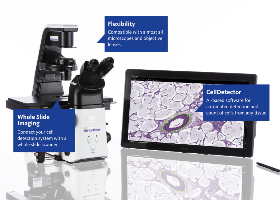
The CellScan multimodal panoramic scanning system is developed by Molecular Machines & Industries (MMI, Germany). It is supplied by default as a complete configuration with an inverted research microscope (Nikon Ti2-E, EVIDENT IX83 / IX85, or ZEISS Axio Observer 7) and converts an entire slide into a high-resolution digital image. Targets can be outlined on the scan using the same microscope and passed to CellCut laser microdissection, so that positional information remains in the coordinate system of the original slide. The system serves spatial multi-omics and related life-science research.

Figure. MMI CellScan on an inverted research microscope.
Positioning and principles
- Defined by the manufacturer as 5D whole-slide imaging: XY scanning, Z-stack, time-lapse imaging, and multicolor fluorescence.
- Sample formats include standard slides, membrane slides for subsequent laser microdissection, and live cells in culture dishes. A stage incubation environment for time-lapse imaging can be configured according to the method; environmental control equipment is not part of the standard system.
- Images are stored in the open BigTIFF format and support remote viewing and annotation.
- The optional CellDetector automated target recognition software assists in locating targets on the whole-slide image before transfer to CellCut.
- The optional ShapeImport data import software imports third-party contours or remote annotations into CellTools before cutting with CellCut. Remote image viewing and contour import are separate functions.
- In China, CellScan is supplied by default as a complete system with one of the host microscopes listed above.
Typical applications
- Whole-slide recording and target outlining for spatial multi-omics and other studies that require in situ tissue coordinates.
- High-resolution whole-slide digitization of tissue sections and membrane slides, with collaborative image review.
- Time-lapse panoramic observation of live cells in culture dishes, with environmental control configured for each project.
- Research workflows linked to laser microdissection on the same microscope. CellScan is not intended as a high-throughput slide scanner for routine pathology laboratories.
Same-microscope workflow with CellCut
- Scan the whole slide to produce a high-resolution digital image.
- Identify and outline targets on the image, either manually, with the assistance of the optional CellDetector software, or by importing external contours with ShapeImport.
- On the same inverted research microscope, CellCut performs laser microdissection with top-side collection and visual confirmation on the cap.
- Positional information is retained in the coordinate system of the original slide, which facilitates comparison with downstream omics results.

Figure. Scan, outline and cut on one optical platform.
Oriental Horizon Technology Co Ltd (Beijing) / 北京欧浩科技有限公司
Authorized distributor in China for Molecular Machines & Industries (MMI, Germany)
Address: Room 2703, Building A, Times Fortune Plaza, No. 1 Hangfeng Road, Fengtai District, Beijing
Demonstrations and evaluations: Ms. Zhang · judy.cheung@ohhktech.com · Mobile +86 135 1102 9964 · Landline 010 81311551
Manufacturer: molecular-machines.com · Chinese site: molecular-machines.cn
For research use only.