


全文约 2150 字(不含文献列表中的英文题录),预计阅读约 6 分钟。
导语
英国 AUROX 公司(Aurox Ltd,位于牛津郡 Abingdon)成立于 2004 年,创始人曾在牛津大学扫描光学显微镜研究组工作。它不使用针孔阵列和激光点扫描,而是用一块刻有条纹图案的旋转玻璃盘做“结构化照明 + 结构化检测”:焦面上的荧光穿过盘面条纹,焦外的模糊光被盘面反射,两路图像同时落在相机的两半,再相减得到光学切片图像(optical section,即只保留焦面附近信号的“薄层”图像)。目前的产品有两条线:Clarity——通过 C 接口加装到现有荧光显微镜上的转盘共聚焦模块;UNITY——集成 LED 光源、双 sCMOS 相机和载物台的桌面式一体机。
我们在 Europe PMC、PubMed Central、Crossref、bioRxiv 等数据库中检索,并逐篇核对了全文 Methods(方法)或图注。本文只收录正文明确写出 Aurox、Clarity 或 UNITY 的论文,仅泛泛提到“转盘共聚焦”而未点名 AUROX 的文章不收录。下面按应用方向介绍代表性工作,每条都附 DOI,便于读者查阅原文。
一、活体钙成像与血管、心肌生理
这一方向的共同需求是:样品活着、信号变化快、光照不能太强。
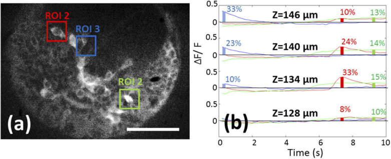
Gintoli 等,Biomedical Optics Express,2020。 作者把 Clarity 转盘与自建的“远程聚焦”光路组合,在不移动样品和物镜的情况下快速切换焦面。他们在 Platynereis(一种海洋环节动物)幼虫神经系统中,以每秒 1 个体积的速度采集 5 个焦面,记录到自发的神经钙信号。DOI: 10.1364/BOE.389904
Lee 等,The Journal of Physiology,2025。 开源血管研究平台 VasoTracker 2 的论文。在加压肠系膜动脉的共聚焦钙成像实验中,作者用 Aurox Clarity LED 结构化照明共聚焦,分别观察平滑肌细胞层和内皮细胞层的钙活动。DOI: 10.1113/JP289322
Deisl 等,Journal of General Physiology,2023。 研究小鼠心肌细胞内肌丝与线粒体造成的纵向扩散屏障,荧光成像在 Nikon Eclipse TE2000 倒置显微镜加装 Aurox Clarity 的系统上完成,物镜为 40× 水镜。DOI: 10.1085/jgp.202213329
O'Reilly 等,Europace,2023。 研究家族性房颤相关的 SCN5A 突变;心脏组织 WGA 染色图像由配备 Aurox Clarity 转盘单元的显微镜和 Visionary 软件采集,配合 Zeiss 20× 物镜。DOI: 10.1093/europace/euac218
Voelkl、Alesutan 团队的血管平滑肌钙化系列(2025–2026)。 三篇论文都使用配有 Clarity 免激光共聚焦单元的显微镜(60× 油镜),研究对象是血管平滑肌细胞钙化的调控因子。DOI: 10.3390/ijms27031411;10.18632/aging.206233;10.1007/s00424-025-03068-6
二、细胞生物学:有丝分裂、线粒体与细胞膜
这类研究多为固定或活细胞的多色成像,看重亚细胞结构的清晰度和日常使用的便利性。UNITY 一体机的已发表应用目前主要集中在这一方向。
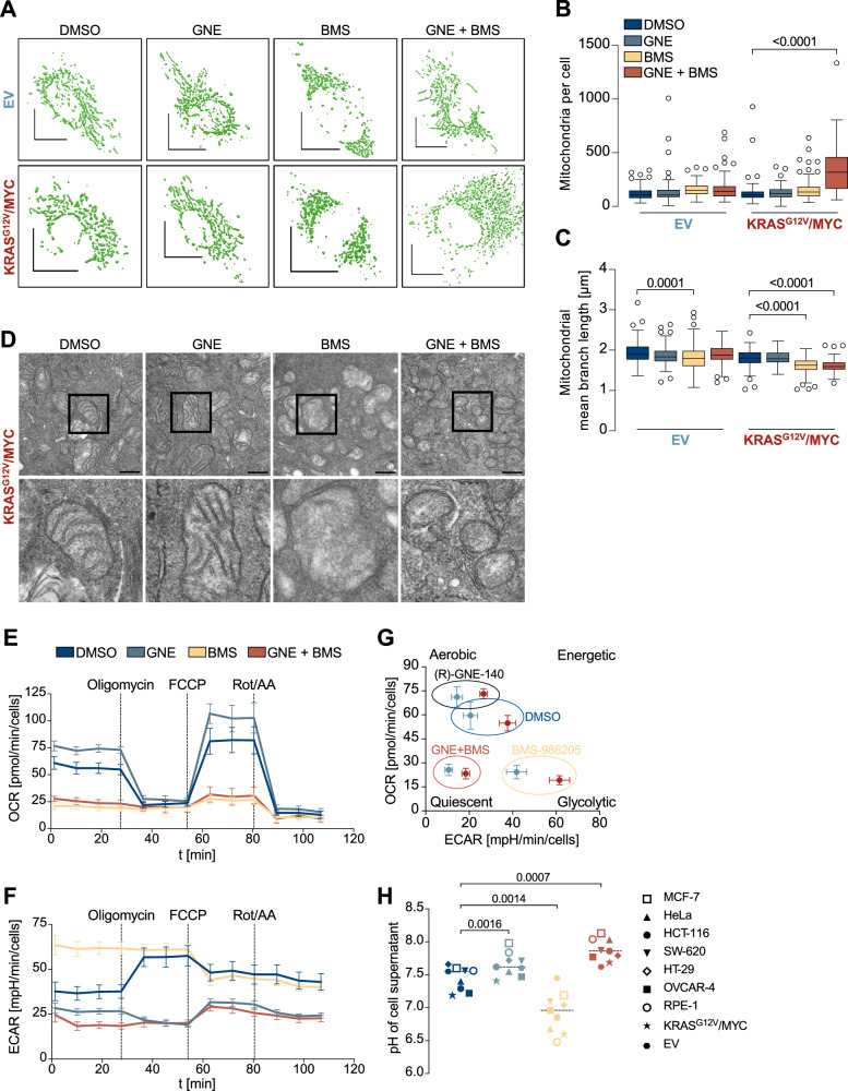
Dreute 等,Cell Death & Differentiation,2025(UNITY)。 作者发现两种代谢抑制剂联用可协同抑制卵巢癌模型细胞,其中 BMS-986205 存在抑制线粒体复合物 I 的脱靶效应。线粒体三维形态和核纤层图像均在 Aurox UNITY 上采集(Nikon 60×/1.4 油镜)。DOI: 10.1038/s41418-025-01532-5
Pfisterer 等,Open Biology,2024(UNITY)。 研究 Aurora B 调控的 PP1/RepoMan 复合物如何决定有丝分裂期组蛋白 H2B S6 磷酸化的时空分布,共聚焦成像在 Aurox UNITY 上完成。DOI: 10.1098/rsob.230460
Deutschmeyer 等,International Journal of Cancer,2025(UNITY)。 研究 SIAH3 在肿瘤中的表观沉默及其对线粒体代谢的调控,共聚焦图像由 Aurox UNITY 采集。DOI: 10.1002/ijc.35202
Deisl 等,Nature Communications,2021。 研究 TMEM16F 与 dynamin 对质膜“储备膜”的调控,成像平台为 Nikon Eclipse 倒置显微镜加装 Clarity。DOI: 10.1038/s41467-021-25286-z
Tikkanen 团队(德国吉森大学)。 研究溶酶体贮积病酶替代疗法(Zeiss Axiovert 200M + Clarity)和琥珀酸半醛脱氢酶缺乏症基因变异(Clarity + Visionary)。DOI: 10.1002/jimd.70248;10.3390/ijms25105237
Kiander 等,Pharmaceutical Research,2021。 研究转运蛋白 OATP1B1 变异体的细胞定位,部分图像在 Leica DM6000 加装 Clarity 的系统上获得。DOI: 10.1007/s11095-021-03107-8
三、发育生物学
果蝇和小鼠的肠道研究常需要大范围拼接和多层组织成像。
Strutynska 等,Development,2026。 研究转录因子 NF-Y 如何通过 mTOR 调控果蝇中肠的营养适应性生长,中肠横切面的多层表型用 Aurox Clarity 成像后定量。DOI: 10.1242/dev.205643
Mattila 等,Science Advances,2024。 研究肠道干细胞 mTOR 信号如何在营养适应中决定区域特异的细胞命运,中肠样品用 Clarity 成像。DOI: 10.1126/sciadv.adi2671
Viitanen 等,Cell Reports Methods,2021。 提出果蝇中肠分区细胞表型分析方法,用 Clarity 对整段中肠做前后方向的拼接扫描。DOI: 10.1016/j.crmeth.2021.100059
Kokki 等,PLoS Genetics,2021。 研究果蝇 GATA 转录因子 Grain 对代谢基因的调控,样品用 Aurox Clarity HS 成像。DOI: 10.1371/journal.pgen.1009855
Marefati 等,Development,2025。 研究小鼠肠道再生中不依赖 Lgr5 的发育谱系,免疫荧光用 Zeiss Axio Observer 加 Aurox 转盘观察。DOI: 10.1242/dev.204654
四、方法学:自适应光学、远程聚焦与开源控制
Hussain 等,Journal of Microscopy,2022。 牛津大学团队在 Clarity 光路中插入可变形镜,实现无波前传感器的自适应光学校正,改善了果蝇神经肌肉接头和卵室样品深处的对比度与细节。DOI: 10.1111/jmi.12976;同一工作的会议版见 Proc. SPIE 2020,DOI: 10.1117/12.2542959
Susano Pinto 等,Journal of Cell Science,2021;Phillips 等,Wellcome Open Research,2021。 两篇开源显微镜控制软件(Python-Microscope、Microscope-Cockpit)论文均以 Aurox Clarity 自适应光学系统为示例平台。DOI: 10.1242/jcs.258955;10.12688/wellcomeopenres.16610.2
Ahmad 等,Frontiers in Robotics and AI,2020。 单细胞荧光分布的图像特征分析研究,示例细胞核图像由 Aurox Clarity 采集。DOI: 10.3389/frobt.2020.00039
远程聚焦方面的工作见上文 Gintoli 2020。
五、感染与病毒入胞
Hudák、Letoha 团队(2021–2023)。 三篇论文分别研究 syndecan 家族在 SARS-CoV-2 入胞中的作用、灭活 SARS-CoV-2 在小鼠体内的分布与内化,以及 Syndecan-4 介导 AAV9 进入细胞。三篇的成像平台都是 Leica DMi8 加装 Aurox Clarity,并用 Visionary 软件采集。DOI: 10.3390/ijms22105336;10.3390/ijms23147609;10.3390/ijms24043141
六、生物材料与干细胞
这类工作常需要在水凝胶、支架等三维材料中观察细胞。
Kiiskinen 等,Stem Cell Research & Therapy,2019;Koivunotko 等,Biomedicines,2022。 研究纳米纤维素敷料和水凝胶中的人间充质干/基质细胞,样品用 Aurox Clarity HS 成像(原文写明为宽场模式)。DOI: 10.1186/s13287-019-1394-7;10.3390/biomedicines10102584
Teixeira Polez 等,Materials Today Bio,2024。 比较纤维素纳米纤丝与 Matrigel 水凝胶中细胞球的形成,用 Zeiss Axio Observer 加装 Clarity 观察。DOI: 10.1016/j.mtbio.2024.101065
Tang 等,J Mater Sci Mater Med,2024。 研究含锌锶的掺钛磷酸盐玻璃用于骨再生,共聚焦成像为 Olympus BX51 正置显微镜 搭载 Aurox,使用 10×、20× 水镜。这是正置机型加装的一个实例。DOI: 10.1007/s10856-024-06804-z
Tan 等,Polymers,2023;Verisqa 等,Biomolecules,2022。 牙周再生水凝胶和 3D 打印骨支架研究,细胞图像用 Aurox 共聚焦与 Visionary 软件采集。DOI: 10.3390/polym15204146;10.3390/biom12111692
七、其他:神经、肿瘤与病理
Nakos Bimpos 等,Acta Neuropathologica Communications,2024。 帕金森病模型研究,用 Aurox Clarity 对脑片做拼接扫描。DOI: 10.1186/s40478-024-01797-w
Thapa 等,Cellular and Molecular Life Sciences,2023。 衰老间皮基质与卵巢癌定植研究,用 Olympus IX83 加装 Aurox 结构化照明转盘。DOI: 10.1007/s00018-023-05017-x
Knudsen 等,Clinical & Experimental Metastasis,2018。 在结直肠癌病理切片上分析 miR-21,共聚焦由 Aurox 免激光结构化照明单元完成。DOI: 10.1007/s10585-018-9945-3
另有皮肤免疫(Zakrzewicz 2022)、食管鳞癌(Moresi 2026)、新生儿糖尿病综合征(Virgilio 2025)、骨关节炎(Jiang 2024、2026)等论文在方法中写明使用 Aurox 转盘共聚焦,详见文末列表。
小结与阅读提示
从已发表文献看,Clarity 的用户覆盖 Leica、Nikon、Olympus/Evident、Zeiss 等品牌的倒置和正置显微镜,应用从细胞器形态、病毒入胞一直延伸到活体钙成像和光学方法开发。UNITY 的论文数量目前较少,但已出现在肿瘤代谢、有丝分裂等细胞生物学研究中。需要说明的是:多数论文只把 AUROX 设备作为成像工具之一,并未与其他共聚焦系统做系统比较;读者引用时应以各论文自身的实验设计和结论为准。
与 AUROX 产品的相关性
Clarity:通过标准 C 接口连接显微镜侧口或顶部相机口。带标准 C 接口的显微镜(包括正置机型)理论上都可以加装,具体型号请提前联系欧浩,由我们与厂家确认适配方案。Clarity 标准版最高 50 fps,HS 版最高 100 fps;默认用 LED 光源,也可以通过扩束器把激光扩成面光源使用。控制软件可选 AUROX Visionary,或 Visitron VisiView(VisiView 支持 Clarity)。
UNITY:桌面一体机,内置 LED 光源与双 sCMOS 相机,适合细胞生物学实验室日常多色共聚焦成像,上文 Dreute、Pfisterer、Deutschmeyer 三篇论文即为实例。
完整文献列表(按本文出现顺序)
Gintoli M, et al. Spinning disk-remote focusing microscopy. Biomed Opt Express. 2020. DOI: 10.1364/BOE.389904(Clarity)
Lee MD, et al. VasoTracker 2: Open-source software and hardware for tracking blood vessel diameter and assessing vascular function. J Physiol. 2025. DOI: 10.1113/JP289322(Clarity)
Deisl C, Chung JH, Hilgemann DW. Longitudinal diffusion barriers imposed by myofilaments and mitochondria in murine cardiac myocytes. J Gen Physiol. 2023. DOI: 10.1085/jgp.202213329(Clarity)
O'Reilly M, et al. Familial atrial fibrillation mutation M1875T-SCN5A increases early sodium current and dampens the effect of flecainide. Europace. 2023. DOI: 10.1093/europace/euac218(Clarity)
Bahiraii S, et al. Role of Inositol Hexakisphosphate Kinases in Vascular Smooth Muscle Cell Calcification. Int J Mol Sci. 2026. DOI: 10.3390/ijms27031411(Clarity)
Razazian M, et al. Fisetin ameliorates vascular smooth muscle cell calcification via DUSP1-dependent p38 MAPK inhibition. Aging (Albany NY). 2025. DOI: 10.18632/aging.206233(Clarity)
Razazian M, et al. Sphingosine kinase 1 inhibition aggravates vascular smooth muscle cell calcification. Pflugers Arch. 2025. DOI: 10.1007/s00424-025-03068-6(Clarity)
Dreute J, et al. Synergistic targeting of cancer cells through simultaneous inhibition of key metabolic enzymes. Cell Death Differ. 2025. DOI: 10.1038/s41418-025-01532-5(UNITY)
Pfisterer M, et al. The Aurora B-controlled PP1/RepoMan complex determines the spatial and temporal distribution of mitotic H2B S6 phosphorylation. Open Biol. 2024. DOI: 10.1098/rsob.230460(UNITY)
Deutschmeyer VE, et al. SIAH3 is frequently epigenetically silenced in cancer and regulates mitochondrial metabolism. Int J Cancer. 2025. DOI: 10.1002/ijc.35202(UNITY)
Deisl C, Hilgemann DW, Syeda R, Fine M. TMEM16F and dynamins control expansive plasma membrane reservoirs. Nat Commun. 2021. DOI: 10.1038/s41467-021-25286-z(Clarity)
Banning A, Reznikova L, Murad A, Tikkanen R. Revisiting Enzyme Replacement Therapy for Aspartylglucosaminuria: Truncated Phosphotransferase Enhances Mannose-6-Phosphorylation and Cellular Uptake of Aspartylglucosaminidase. J Inherit Metab Dis. 2026. DOI: 10.1002/jimd.70248(Clarity)
Didiasova M, et al. Functional Characterization of a Spectrum of Genetic Variants in a Family with Succinic Semialdehyde Dehydrogenase Deficiency. Int J Mol Sci. 2024. DOI: 10.3390/ijms25105237(Clarity)
Kiander W, et al. The Effect of Single Nucleotide Variations in the Transmembrane Domain of OATP1B1 on in vitro Functionality. Pharm Res. 2021. DOI: 10.1007/s11095-021-03107-8(Clarity)
Strutynska T, Deniz O, Mattila J. Nuclear Factor Y controls nutrient-adaptive epithelial growth by regulating mTOR in the Drosophila midgut. Development. 2026. DOI: 10.1242/dev.205643(Clarity)
Mattila J, et al. Stem cell mTOR signaling directs region-specific cell fate decisions during intestinal nutrient adaptation. Sci Adv. 2024. DOI: 10.1126/sciadv.adi2671(Clarity)
Viitanen A, et al. An image analysis method for regionally defined cellular phenotyping of the Drosophila midgut. Cell Rep Methods. 2021. DOI: 10.1016/j.crmeth.2021.100059(Clarity)
Kokki K, et al. Metabolic gene regulation by Drosophila GATA transcription factor Grain. PLoS Genet. 2021. DOI: 10.1371/journal.pgen.1009855(Clarity HS)
Marefati M, et al. A Lgr5-independent developmental lineage is involved in mouse intestinal regeneration. Development. 2025. DOI: 10.1242/dev.204654(Aurox 转盘)
Hussain SA, et al. Wavefront-sensorless adaptive optics with a laser-free spinning disk confocal microscope. J Microsc. 2022. DOI: 10.1111/jmi.12976(Clarity)
Hussain SA, et al. Sensorless adaptive optics with a laser free spinning disk confocal microscope. Proc SPIE 11248. 2020. DOI: 10.1117/12.2542959(Clarity)
Susano Pinto DM, et al. Python-Microscope – a new open-source Python library for the control of microscopes. J Cell Sci. 2021. DOI: 10.1242/jcs.258955(Clarity)
Phillips MA, et al. Microscope-Cockpit: Python-based bespoke microscopy for bio-medical science. Wellcome Open Res. 2021. DOI: 10.12688/wellcomeopenres.16610.2(Clarity)
Ahmad A, Frindel C, Rousseau D. Detecting Differences of Fluorescent Markers Distribution in Single Cell Microscopy: Textural or Pointillist Feature Space? Front Robot AI. 2020. DOI: 10.3389/frobt.2020.00039(Clarity)
Hudák A, Letoha A, Szilák L, Letoha T. Contribution of Syndecans to the Cellular Entry of SARS-CoV-2. Int J Mol Sci. 2021. DOI: 10.3390/ijms22105336(Clarity)
Hudák A, et al. Biodistribution and Cellular Internalization of Inactivated SARS-CoV-2 in Wild-Type Mice. Int J Mol Sci. 2022. DOI: 10.3390/ijms23147609(Clarity)
Hudák A, et al. Syndecan-4 Mediates the Cellular Entry of Adeno-Associated Virus 9. Int J Mol Sci. 2023. DOI: 10.3390/ijms24043141(Clarity)
Kiiskinen J, et al. Nanofibrillar cellulose wound dressing supports the growth and characteristics of human mesenchymal stem/stromal cells without cell adhesion coatings. Stem Cell Res Ther. 2019. DOI: 10.1186/s13287-019-1394-7(Clarity HS)
Koivunotko E, et al. Angiogenic Potential of Human Adipose-Derived Mesenchymal Stromal Cells in Nanofibrillated Cellulose Hydrogel. Biomedicines. 2022. DOI: 10.3390/biomedicines10102584(Clarity HS)
Teixeira Polez R, et al. Insights into spheroids formation in cellulose nanofibrils and Matrigel hydrogels using AFM-based techniques. Mater Today Bio. 2024. DOI: 10.1016/j.mtbio.2024.101065(Clarity)
Tang T, et al. Titanium-doped phosphate glasses containing zinc and strontium applied in bone regeneration. J Mater Sci Mater Med. 2024. DOI: 10.1007/s10856-024-06804-z(Aurox,Olympus BX51)
Tan N, et al. β-Tricalcium Phosphate-Loaded Chitosan-Based Thermosensitive Hydrogel for Periodontal Regeneration. Polymers. 2023. DOI: 10.3390/polym15204146(Aurox + Visionary)
Verisqa F, et al. Digital Light Processing 3D Printing of Gyroid Scaffold with Isosorbide-Based Photopolymer for Bone Tissue Engineering. Biomolecules. 2022. DOI: 10.3390/biom12111692(Aurox + Visionary)
Nakos Bimpos M, et al. Alpha-synuclein-induced stress sensitivity renders the Parkinson's disease brain susceptible to neurodegeneration. Acta Neuropathol Commun. 2024. DOI: 10.1186/s40478-024-01797-w(Clarity)
Thapa BV, Banerjee M, Glimm T, Saini DK, Bhat R. The senescent mesothelial matrix accentuates colonization by ovarian cancer cells. Cell Mol Life Sci. 2023. DOI: 10.1007/s00018-023-05017-x(Aurox 转盘)
Knudsen KN, et al. miR-21 expression analysis in budding colon cancer cells by confocal slide scanning microscopy. Clin Exp Metastasis. 2018. DOI: 10.1007/s10585-018-9945-3(Aurox 结构化照明单元)
Zakrzewicz A, et al. Stabilization of Keratinocyte Monolayer Integrity in the Presence of Anti-Desmoglein-3 Antibodies through FcRn Blockade with Efgartigimod. Cells. 2022. DOI: 10.3390/cells11060942(Clarity)
Moresi F, et al. CDK4/6 inhibition uncovers subtype-specific vulnerabilities and immune-related responses in esophageal squamous cell carcinoma. Cell Death Dis. 2026. DOI: 10.1038/s41419-026-08892-x(Aurox LFC)
Virgilio E, et al. Recessive TMEM167A variants cause neonatal diabetes, microcephaly, and epilepsy syndrome. J Clin Invest. 2025. DOI: 10.1172/jci195756(Aurox 转盘)
Jiang S, et al. Transcript-dependent effects of the CALCA gene on the progression of post-traumatic osteoarthritis in mice. Commun Biol. 2024. DOI: 10.1038/s42003-024-05889-0(Aurox 转盘)
Jiang S, et al. The calcitonin receptor controls osteophyte formation, but not cartilage degeneration and subchondral bone loss in experimental osteoarthritis. Bone Joint Res. 2026. DOI: 10.1302/2046-3758.152.bjr-2025-0025.r1(Aurox 转盘)
说明:本文为对上述公开文献的整理导读,非各论文作者的官方译文;设备信息摘自各论文 Methods 或图注,检索截至 2026 年 10 月。
北京欧浩科技有限公司
邮箱:lili@ohhktech.com
AUROX 中文站:https://www.aurox.com.cn